Advanced Technology
All technologies used at Queen City Endodontics are carefully chosen to make the patient experience better, while helping us to be more efficient and productive. From our fully digital/paperless office to digital 2D and more advanced focused 3D imaging (CBCT), our patients learn just how big of a difference the right technology can make. Here are some examples of the technology that we use at Queen City Endodontics to help us deliver the exceptional endodontic care we promise for all of our patients.
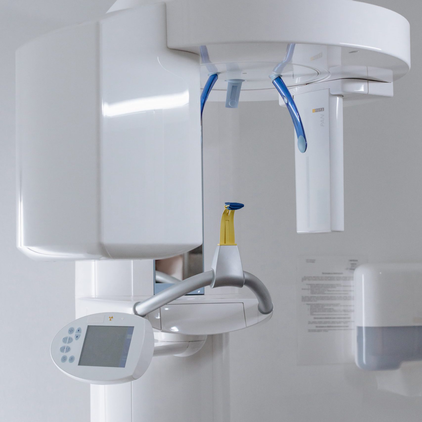
CBCT 3D Imaging
3D imaging (CBCT) is an advanced dental imaging technology that allows for a 3-dimensional, slice by slice, view of the teeth and supportive structures. The CBCT can capture panoramic views of the maxillofacial area in just 10 seconds. It offers an enhanced view of the face that can be examined and evaluated from multiple viewing angles. A single image can reveal the associative relationship between the hard and soft tissues of the mouth, which aid in diagnosis and treatment planning. These 3D images allow diagnosis of infections that cannot be seen on 2D images.
CBCTs are similar to the images available through CT scans. The difference is that they use far less radiation, takes less time, and the images can easily be obtained in a dental office. CBCT images are stored digitally for future reference or for easy transfer between providers. The scan is completely painless and there is no pre-scan consultation or after care instructions required.

Surgical Operating Microscope
A surgical operating microscope is used for every treatment. This provides the magnification and illumination needed to visualize the minutest details inside your tooth. Proper endodontic therapy is much more difficult, to perform without the use of the operating microscope.
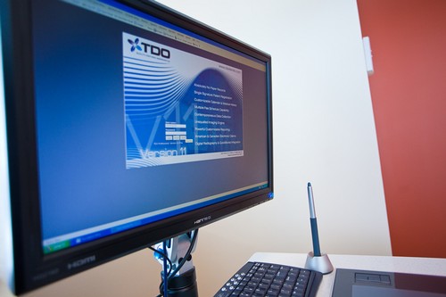
TDO Software
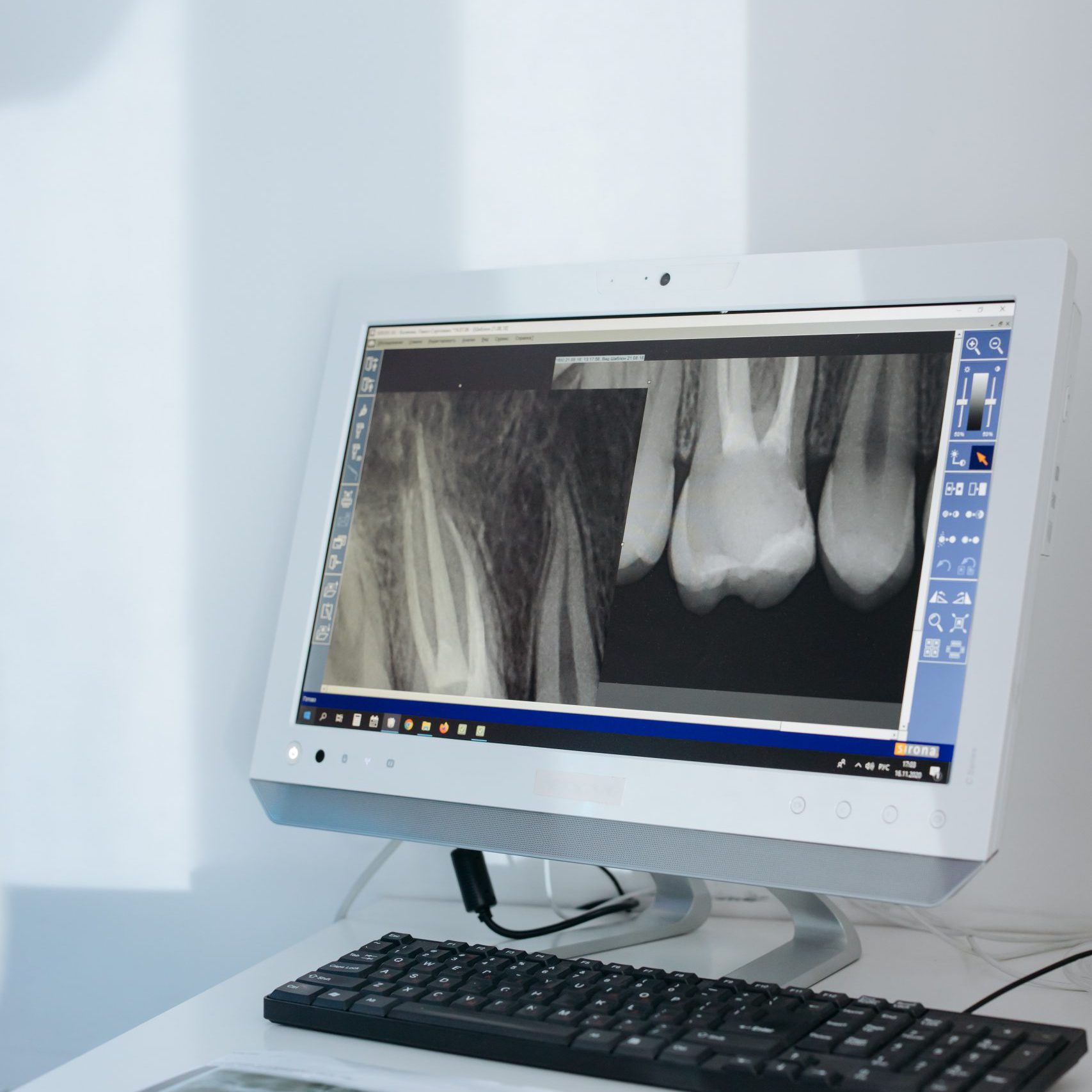
Digital Radiography
Digital x-rays are a more streamlined way of taking dental radiographs. Like traditional x-rays, digital versions provide an in-depth view of the structures of the mouth, helping detect complications and develop effective methods of treatment. Digital x-rays are capable of revealing hidden tooth decay and bone recession.
Requiring less radiation and no film to process, digital x-rays have become the standard for oral imaging. These systems produce instant digital images that can easily be enhanced and enlarged for a more accurate diagnosis. The images are captured, stored, and even transmitted via in-office computers. They can be easily printed or emailed in just seconds.
Digital x-rays make for a better and more efficient patient experience. Office visits are faster, patients are exposed to less radiation, and radiographs can be sent to other specialists for review in a fraction of the time necessary for traditional film x-rays.